

Matthew Pellicore’s Qualifying Exam was titled "Toward understanding the role of exosome-mediated paracrine signaling in the degradation of cartilage in the osteoarthritic joint."
Congratulations, Matt!
Matthew Pellicore’s Qualifying Exam was titled "Toward understanding the role of exosome-mediated paracrine signaling in the degradation of cartilage in the osteoarthritic joint."
Congratulations, Matt!
Dr. Clark Hung presented the lab’s recent research at Tissue Talks hosted by Dr. Gordana Vunjak-Novakovic and the Laboratory for Stem Cells and Tissue Engineering.
The subject of the talk was “The synovium role in joint inflammation and repair.”
Andy Lee was awarded an NIH F31 fellowship for his project titled “Ferroptosis as a Potential Mechanism of Blood-Induced Chondrocyte Cell Death.”
Congratulations, Andy!
Justin Hellwinkel, an Orthopedic Surgery Resident at Columbia Presbyterian Hospital, will be co-advised by Dr. David Trofa and Dr. Clark Hung.
Welcome back, Sofía!

Lance Murphy, former CEL undergrad and technician, joins the University of Pennsylvania Medical Scientist Training Program (MSTP).
Congratulations and good luck on your MD-PhD journey, Lance!
Lianna Gangi received Honorable Mention on her 2020 National Science Foundation Graduate Research Fellowship Program (NSF-GRFP) proposal titled Bioengineering model of synovium to study osteoarthritic pain. Congratulations, Lianna!
We would also like to congratulate fellow BME graduate students Ethan Bendau and McKenzie Sup!

Jack Rogot won 3rd place at the Poster Competition during the Engineering in Medicine Symposium held at Columbia University on February 20, 2020.

Articular cartilage injuries are a common source of joint pain and dysfunction. We hypothesized that pulsed electromagnetic fields (PEMFs) would improve growth and healing of tissue engineered cartilage grafts in a time‐ and direction‐dependent manner. PEMF stimulation of engineered cartilage constructs was first evaluated in vitro using passaged adult canine chondrocytes embedded in an agarose hydrogel scaffold. PEMF coils oriented parallel to the articular surface induced superior repair stiffness compared to both perpendicular PEMF (p=0.026) and control (p=0.012). This was correlated with increased GAG deposition in both parallel and perpendicular PEMF orientations compared to control (p=0.010 and 0.028, respectively). Following in vitro optimization, the potential clinical translation of PEMF was evaluated in a preliminary in vivo preclinical adult canine model. Engineered osteochondral constructs (∅ 6 mm x 6 mm thick, devitalized bone base) were cultured to maturity and implanted into focal defects created in the stifle (knee) joint. To assess expedited early repair, animals were assessed after a 3‐month recovery period, with microfracture repairs serving as an additional clinical control. In vivo, PEMF led to a greater likelihood of normal chondrocyte (OR: 2.5, p=0.051) and proteoglycan (OR: 5.0, p=0.013) histological scores in engineered constructs. Interestingly, engineered constructs outperformed microfracture in clinical scoring, regardless of PEMF treatment (p<0.05). Overall, the studies provided evidence that PEMF stimulation enhanced engineered cartilage growth and repair, demonstrating a potential low‐cost, low‐risk, non‐invasive treatment modality for expediting early cartilage repair.
Articular cartilage defects are a common source of joint pain and dysfunction. We hypothesized that sustained low-dose dexamethasone (DEX) delivery via an acellular osteochondral implant would have a dual pro-anabolic and anti-catabolic effect, both supporting the functional integrity of adjacent graft and host tissue while also attenuating inflammation caused by iatrogenic injury. An acellular agarose hydrogel carrier with embedded DEX-loaded poly(lactic-co-glycolic) acid (PLGA) microspheres (DLMS) was developed to provide sustained release for at least 99 days. The DLMS implant was first evaluated in an in vitro pro-inflammatory model of cartilage degradation. The implant was chondroprotective, as indicated by maintenance of Young's modulus (EY) (p=0.92) and GAG content (p=1.0) in the presence of interleukin-1β insult. In a subsequent preliminary in vivo experiment, an osteochondral autograft transfer was performed using a pre-clinical canine model. DLMS implants were press-fit into the autograft donor site and compared to intra-articular DEX injection (INJ) or no DEX (CTL). Functional scores for DLMS animals returned to baseline (p=0.39), whereas CTL and INJ remained significantly worse at 6 months (p<0.05). DLMS knees were significantly more likely to have improved OARSI scores for proteoglycan, chondrocyte, and collagen pathology (p<0.05). However, no significant improvements in synovial fluid cytokine content were observed. In conclusion, utilizing a targeted DLMS implant, we observed in vitro chondroprotection in the presence of IL-1-induced degradation and improved in vivo functional outcomes. These improved outcomes were correlated with superior histological scores but not necessarily a dampened inflammatory response, suggesting a primarily pro-anabolic effect.
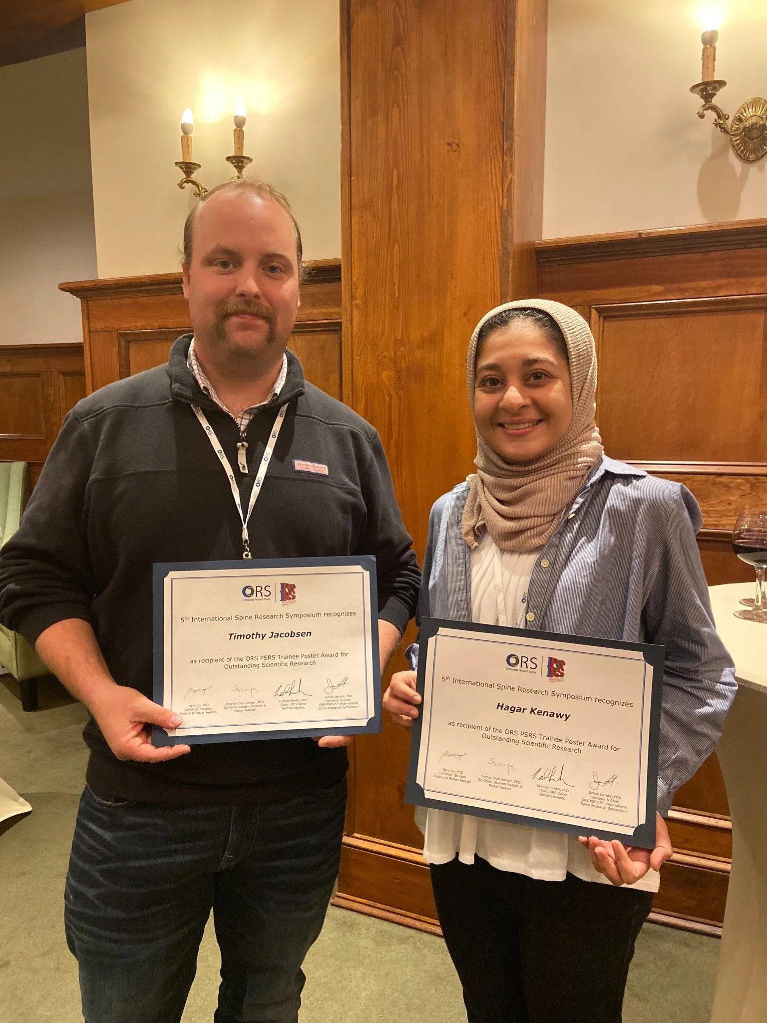
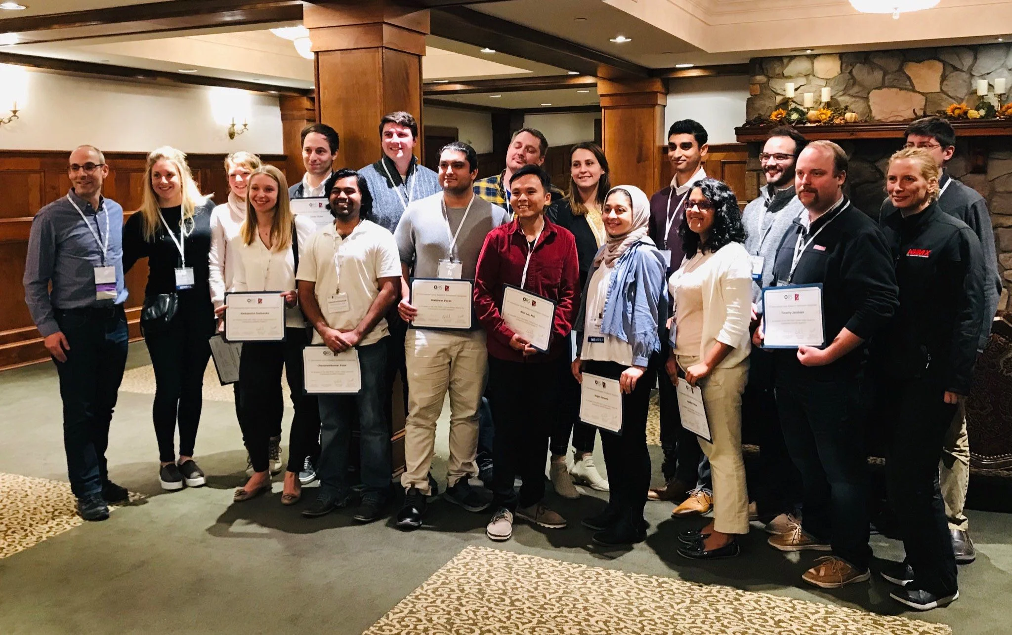
Hagar Kenawy (CEL, Columbia University) and Timothy Jacobsen (Chahine Lab, Columbia University) received the ORS PSRS Trainee Poster Award for Outstanding Scientific Research at the 5th International Spine Research Symposium in Skytop, Pennsylvania. Congratulations!